


Technology -
Scanning Laser Confocal Microscope
Many people will remember their experience with microscopes.
Even though the object was magnified, certain details always appeared to be
fuzzy. At work is a basic principle of optics that prevents both
magnification and the ability to view all the details of a thick specimen.
The term used to describe the thickness of the sample that can be viewed in
sharp detail is called depth-of-field. Up to the mid 1980's depth-of-field
limited the ability to see objects in detail. This is especially true at high
magnifications. Early in the 1980's inexpensive lasers, high speed computers,
and a very ingenious microscope design were combined to create the first
commercial scanning-laser confocal microscope.
Confocal imaging - what is it?
The basic principle of a confocal microscope is shown below.
 In the
confocal microscope all structures that are out of
focus are eliminated at image formation. In the
confocal microscope all structures that are out of
focus are eliminated at image formation.
This is possible because the object is not being
illuminated and imaged entirely at the same time, but rather one
single point after the other. As shown
in the diagram, this is obtained by the
arrangement of diaphragms, which act as a
point source and as a point detector respectively (solid line). Rays from out-of-focus
areas are prevented from being imaged by the final confocal pinhole opening (dashed
lines).
Using a confocal microscope eliminates out of focus points
while retaining the sharp sample areas. Part of the problem for microscopes
has been solved. Depth-of-field has been circumvented and sharp images can be
viewed, but only for very thin slices.
What about thick materials? The next important
development was high speed inexpensive graphic computers and 3-D software.
-
Using a computer, multiple slices of in-focus images are
collected and saved. These slices as a group are called a stack.
-
Using 3-D software, the stack is reassembled into one
sharp image (also called a projection). Take a look and see how
this works.
What does a confocal microscope look like?
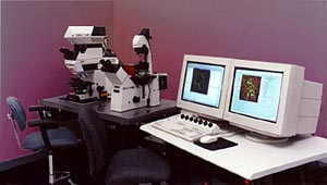 The Leica TCS NT is a universal Laser Scanning Confocal Microscope
System for the bio-medical and materials research environment. The system includes
four lasers that can be used simultaneously. The lasers and laser lines include,
an Argon (488nm), Krypton (568), RHeNe (633), and an Argon UV (363).
The Leica TCS NT is a universal Laser Scanning Confocal Microscope
System for the bio-medical and materials research environment. The system includes
four lasers that can be used simultaneously. The lasers and laser lines include,
an Argon (488nm), Krypton (568), RHeNe (633), and an Argon UV (363).
There are two separate microscopes (upright and inverted) that can
accommodate the scanning head, which makes the system flexible for
various research needs.
What are the advantages and limitations of confocal microscopes?
Major improvements offered by a confocal microscope over
the performance of a conventional microscope may be
summarized as follows:
- Light rays from outside the focal plane will not be
recorded.
- Defocusing does not create blurring, but gradually
cuts
out parts of the object as they move away from the
focal plane. The practical consequence is that these
parts become darker and eventually disappear. This
feature is called optical sectioning.
- True, three-dimensional data sets can be recorded.
- Scanning the object in x/y-direction as well as in
z-direction (along the optical axis) allows viewing the
object from all sides.
- Due to the small dimension of the illuminating light
spot in the focal plane, stray light is minimized.
- By image processing, many slices can be superimposed,
giving an extended focus image which can only be achieved
in conventional microscopy by reduction of the aperture
and thus sacrificing resolution.
|
